The vestibular system plays a vital role in our ability to maintain balance and a sense of spatial orientation. Integral to this system is the vestibular nerve, which transmits signals from the inner ear to the brain. In order to understand the functioning of this nerve, it is important to explore its anatomy and function.
Understanding the Vestibular Nerve
The vestibular nerve is a fascinating component of the human body that plays a crucial role in our sense of balance and spatial orientation. It is one of the two branches of the vestibulocochlear nerve, working in tandem with the cochlear nerve to ensure proper functioning of the inner ear.
Anatomy of the Vestibular Nerve
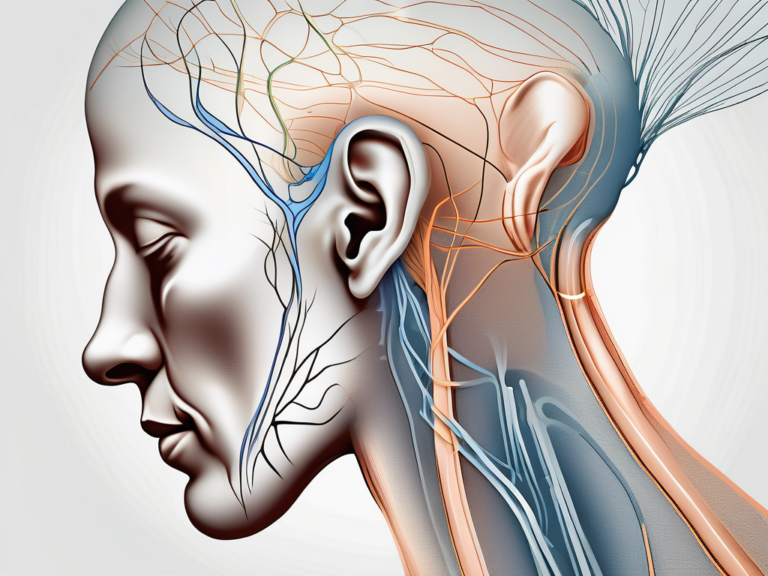
Let’s delve deeper into the anatomy of the vestibular nerve. It originates from the vestibular ganglion, a sensory ganglion located in the internal acoustic meatus of the inner ear. This ganglion serves as the starting point for the vestibular nerve, which consists of two distinct branches – the superior and inferior branches.
The superior branch of the vestibular nerve arises from the utricle and the superior semicircular canal. These structures are essential for detecting linear acceleration and head movements in the vertical plane. On the other hand, the inferior branch of the vestibular nerve originates from the saccule and the posterior semicircular canal. These structures are responsible for sensing head movements in the horizontal plane.
Now that we understand the origins of the vestibular nerve, let’s explore its composition. The fibers of the vestibular nerve are primarily composed of afferent neurons. These neurons play a vital role in transmitting signals from the peripheral receptor cells in the vestibular apparatus to the central nervous system.
As the fibers of the vestibular nerve travel through the internal auditory canal, they embark on a journey to relay crucial information to the brain. Once they reach the brainstem, they synapse with the vestibular nuclei. This synapse is a critical step in the transmission of sensory information, as it allows for further processing and integration with other sensory inputs.
Function of the Vestibular Nerve
The vestibular nerve serves a fundamental function in our daily lives – relaying sensory information about head position, movement, and spatial orientation to the brain. This information is crucial for maintaining balance, coordinating eye movements, and modulating muscle tone.
Imagine walking on a narrow balance beam or riding a roller coaster. These activities require precise coordination of movements, and the vestibular nerve plays a pivotal role in ensuring that we can navigate such challenges with ease. By providing continuous feedback to the brain, the vestibular nerve helps us adjust our body position and muscle tone, allowing us to maintain balance and stability.
Disorders of the vestibular nerve can have a significant impact on an individual’s daily life. Symptoms such as vertigo, dizziness, and imbalance can arise, making simple tasks challenging and potentially affecting overall well-being. If you are experiencing persistent or concerning symptoms related to the vestibular nerve, it is advisable to consult with a medical professional for a thorough evaluation and appropriate management.
In conclusion, the vestibular nerve is a remarkable component of our sensory system. Its intricate anatomy and crucial function in maintaining balance and spatial orientation make it an essential part of our everyday lives. Understanding the vestibular nerve can help us appreciate the complexity of our bodies and seek appropriate care when needed.
The Sensory Ganglion: An Overview
The sensory ganglion is a fascinating component of the nervous system that plays a crucial role in sensory perception. It is a collection of cell bodies of sensory neurons located outside the central nervous system, acting as a relay station for transmitting signals from the peripheral sensory organs to the brain.
These ganglia are associated with various cranial and spinal nerves, each serving a specific sensory function. They are responsible for relaying information related to touch, temperature, pain, proprioception, and other sensory modalities.
Defining Sensory Ganglion
A sensory ganglion is a specialized cluster of cell bodies that are primarily found in the peripheral nervous system. These ganglia are strategically positioned along the pathways of sensory nerves, allowing for efficient transmission of sensory information to the brain.
Each sensory ganglion consists of numerous sensory neurons, which are specialized cells capable of detecting and transmitting sensory signals. These neurons have long, slender projections called axons that extend from the ganglion to the peripheral sensory organs, such as the skin, muscles, and internal organs.
Within the sensory ganglion, the cell bodies of these sensory neurons are densely packed together, forming a distinct structure. This arrangement allows for efficient communication and processing of sensory information before it is relayed to the central nervous system.
Role and Importance of Sensory Ganglion
One of the most well-known sensory ganglia is the vestibular ganglion, also known as Scarpa’s ganglion. This ganglion is associated with the vestibular nerve, which is responsible for transmitting information related to balance and spatial orientation.
The vestibular ganglion, named after the Italian anatomist Antonio Scarpa, contains the cell bodies of afferent neurons that receive signals from the vestibular organs in the inner ear. These organs, known as the semicircular canals and the otolith organs, detect changes in head position and movement.
When the head moves, the sensory receptors within the vestibular organs are stimulated, and this information is transmitted to the vestibular ganglion. From there, the sensory signals are relayed to the brain, specifically to the vestibular nuclei in the brainstem, where they are processed and integrated with other sensory inputs.
The role of the vestibular ganglion is crucial in maintaining balance and coordinating movements. It allows us to perceive our body’s position in space, adjust our posture, and make precise movements with accuracy.
In addition to the vestibular ganglion, there are numerous other sensory ganglia throughout the body, each with its specific function and importance. These ganglia enable us to experience and interpret the world around us, providing us with a rich sensory experience.
Overall, the sensory ganglion is a remarkable structure that plays a vital role in sensory perception. It acts as a gateway, allowing sensory information to be transmitted from the peripheral sensory organs to the brain, ultimately shaping our perception of the world.
The Association between the Vestibular Nerve and Sensory Ganglion
How the Vestibular Nerve and Sensory Ganglion Interact
The vestibular nerve and the sensory ganglion are closely interconnected in the transmission of sensory information. The peripheral processes of the vestibular ganglion neurons extend into the vestibular apparatus of the inner ear, where they detect mechanical stimuli associated with head movements. These stimuli are then converted into electrical signals, which are transmitted along the fibers of the vestibular nerve to the sensory ganglion.
Once the electrical signals reach the sensory ganglion, a complex series of interactions take place. The ganglion acts as a central hub for processing and integrating the incoming signals. Specialized cells within the ganglion, known as ganglion cells, receive and analyze the electrical signals, extracting important information about the direction, speed, and intensity of head movements.
After the signals are processed within the sensory ganglion, they are ready to be relayed to the brainstem. The vestibular nuclei, located in the brainstem, receive input from both sides of the brain and play a crucial role in coordinating the appropriate motor responses to maintain balance and spatial orientation.
The intricate interaction between the vestibular nerve and sensory ganglion ensures the seamless transmission of sensory information from the inner ear to the brain. This coordinated process allows us to perceive and respond to changes in our spatial environment with remarkable precision.
Impact of this Association on Sensory Perception
The association between the vestibular nerve and sensory ganglion plays a crucial role in our ability to perceive and respond to changes in our spatial environment. By detecting head movements and position, the vestibular system contributes to our sense of equilibrium and allows us to navigate the world safely and effectively.
Imagine walking on a narrow bridge or riding a roller coaster. Without the precise coordination between the vestibular nerve and sensory ganglion, our ability to maintain balance and adjust our body position would be severely compromised. The information transmitted through this association helps us make split-second adjustments to keep ourselves upright and stable.
Disruptions in the functioning of the vestibular nerve or sensory ganglion can lead to vestibular disorders, which often manifest as problems with balance and coordination. These disorders can have a significant impact on an individual’s quality of life, affecting their ability to perform everyday tasks and engage in activities they once enjoyed.
It is essential for individuals experiencing symptoms such as dizziness, vertigo, or unsteadiness to seek medical attention. A healthcare professional can evaluate the symptoms, perform diagnostic tests, and develop a personalized treatment plan to manage the vestibular disorder effectively.
In conclusion, the association between the vestibular nerve and sensory ganglion is a remarkable example of the intricate mechanisms that underlie our sensory perception and motor coordination. Understanding the role of this association can help us appreciate the complexity of our own physiological systems and the importance of maintaining their proper functioning.
Identifying the Sensory Ganglion of the Vestibular Nerve
Characteristics of the Vestibular Nerve’s Sensory Ganglion
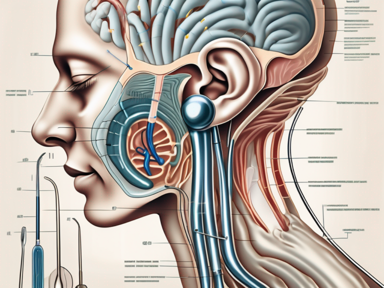
The vestibular ganglion, or Scarpa’s ganglion, is located within the bony labyrinth of the inner ear, specifically in the internal acoustic meatus. It is a small, rounded structure containing the cell bodies of the vestibular nerve’s afferent neurons. These neurons have specialized processes that detect the mechanical stimuli associated with head movements.
The vestibular ganglion is anatomically distinct from the cochlear ganglion, which contains the cell bodies of the cochlear nerve’s afferent neurons responsible for auditory perception. While the two ganglia are closely connected, each serves a distinct sensory modality.
The vestibular ganglion plays a crucial role in our perception of balance and spatial orientation. It is responsible for detecting and transmitting information about head movements and position to the brain. This information is essential for maintaining equilibrium and coordinating our body’s response to changes in our environment.
Within the vestibular ganglion, the cell bodies of the vestibular nerve’s afferent neurons are organized into distinct groups or clusters. These clusters correspond to the different functional subdivisions of the vestibular apparatus, which include the semicircular canals and the otolith organs. Each cluster of neurons within the ganglion is specialized to detect specific types of head movements and provide precise information about our body’s position in space.
The vestibular ganglion is richly supplied with blood vessels, ensuring a constant supply of oxygen and nutrients to support the metabolic needs of the ganglion cells. Additionally, the ganglion is surrounded by a protective layer of connective tissue, which helps to maintain the structural integrity of the ganglion and prevent damage from mechanical forces.
Importance of the Vestibular Nerve’s Sensory Ganglion in the Human Body
The sensory ganglion associated with the vestibular nerve is essential for our sense of balance and spatial orientation. It acts as a relay station, transmitting information from the peripheral vestibular apparatus to the central nervous system. Without the proper functioning of this ganglion, our ability to maintain equilibrium and respond appropriately to changes in our environment would be compromised.
When we move our head, the vestibular ganglion detects the motion and sends signals to the brain, which then processes this information and generates appropriate motor responses to maintain balance. For example, if we suddenly tilt our head to one side, the vestibular ganglion detects the change in head position and sends signals to the brain, which in turn activates the muscles in our neck and body to counteract the tilt and keep us upright.
In addition to maintaining balance, the vestibular ganglion also plays a role in coordinating eye movements. When we move our head, the vestibular ganglion sends signals to the brain, which then adjusts the position of our eyes to compensate for the head movement. This ensures that our visual field remains stable and clear, even when our head is in motion.
Furthermore, the vestibular ganglion is involved in the perception of motion and spatial orientation. It provides the brain with information about the direction and speed of our head movements, allowing us to navigate our environment and interact with objects accurately. This information is crucial for activities such as walking, running, and driving, where precise spatial awareness is essential for safety and coordination.
In summary, the sensory ganglion of the vestibular nerve, located within the inner ear, is a vital component of our sensory system. It enables us to maintain balance, coordinate our movements, and perceive our position in space. Without the proper functioning of this ganglion, our ability to navigate the world around us would be severely impaired.
Disorders Related to the Vestibular Nerve’s Sensory Ganglion
Common Disorders and Their Symptoms
Disorders affecting the vestibular nerve’s sensory ganglion can lead to a range of symptoms and conditions. Some common vestibular disorders include benign paroxysmal positional vertigo (BPPV), Meniere’s disease, vestibular neuritis, and labyrinthitis.
BPPV typically presents with brief episodes of intense vertigo triggered by specific head movements. This condition occurs when tiny calcium crystals in the inner ear become dislodged and migrate into the semicircular canals, causing abnormal fluid movement and triggering vertigo. The vertigo experienced during BPPV can be so severe that it leads to nausea and vomiting. However, the episodes are usually short-lived and can be managed with specific head positioning maneuvers.
Meniere’s disease is characterized by episodes of vertigo, fluctuating hearing loss, tinnitus, and a feeling of fullness in the affected ear. This chronic condition is believed to be caused by an abnormal buildup of fluid in the inner ear, leading to increased pressure and disruption of the delicate balance mechanisms. The vertigo experienced during Meniere’s disease can be debilitating and may last for several hours. Treatment options for Meniere’s disease include medication to manage symptoms, dietary changes to reduce fluid retention, and in severe cases, surgical interventions to alleviate pressure in the inner ear.
Vestibular neuritis and labyrinthitis are inflammatory conditions that can cause severe vertigo, dizziness, and imbalance. Vestibular neuritis typically occurs due to a viral infection that affects the vestibular nerve, leading to inflammation and disruption of normal vestibular function. Labyrinthitis, on the other hand, involves inflammation of the entire inner ear, affecting both the vestibular and cochlear systems. In addition to vertigo, individuals with vestibular neuritis or labyrinthitis may experience hearing loss, ear pain, and a sensation of pressure in the affected ear. Treatment for these conditions focuses on managing symptoms, reducing inflammation, and promoting vestibular compensation through exercises and physical therapy.
Treatment and Management of These Disorders
The treatment and management of vestibular disorders associated with the sensory ganglion of the vestibular nerve require a thorough evaluation by a healthcare professional. Depending on the specific condition and its severity, various treatment approaches may be recommended. These can include medications, vestibular rehabilitation therapy, lifestyle modifications, and in some cases, surgical interventions.
Medications commonly used to manage vestibular disorders include anti-vertigo medications, such as antihistamines and benzodiazepines, which help alleviate symptoms of dizziness and vertigo. Additionally, medications that target the underlying cause of the disorder, such as antiviral drugs for vestibular neuritis, may be prescribed.
Vestibular rehabilitation therapy (VRT) is a specialized form of physical therapy that focuses on improving balance and reducing symptoms of dizziness and vertigo. VRT involves a series of exercises and maneuvers designed to promote vestibular compensation, enhance gaze stability, and improve postural control. This therapy is typically tailored to the individual’s specific needs and can be highly effective in managing vestibular disorders.
Lifestyle modifications can also play a significant role in the management of vestibular disorders. These may include dietary changes, such as reducing salt intake to minimize fluid retention in Meniere’s disease, and avoiding triggers that worsen symptoms, such as certain head movements in BPPV. Stress management techniques, regular exercise, and adequate sleep can also contribute to overall well-being and symptom reduction.
In some cases, surgical interventions may be necessary to alleviate symptoms and improve quality of life. Surgical options for vestibular disorders can vary depending on the specific condition and its underlying cause. For example, in severe cases of Meniere’s disease that do not respond to conservative treatments, a procedure called endolymphatic sac decompression may be performed to reduce fluid pressure in the inner ear. Other surgical interventions may involve removing or repairing damaged structures in the inner ear or implanting devices to stimulate the vestibular system.
It is crucial to consult with a medical professional experienced in vestibular disorders to receive appropriate diagnosis, treatment, and management strategies tailored to your individual needs. Self-diagnosis and self-treatment can lead to ineffective outcomes and potentially exacerbate symptoms. Seeking professional guidance ensures that you receive comprehensive care and support throughout your vestibular disorder journey.
In conclusion, the sensory ganglion associated with the vestibular nerve is commonly known as the vestibular ganglion or Scarpa’s ganglion. This ganglion plays a crucial role in relaying sensory information related to balance and spatial orientation from the peripheral vestibular apparatus to the brain. Disruptions within this complex network can result in various vestibular disorders, which can significantly affect an individual’s well-being. If you are experiencing symptoms or concerns related to your vestibular function, it is advisable to consult with a medical professional for a comprehensive assessment and appropriate management.



+ There are no comments
Add yours