The human body is a marvel of complex structures and intricate connections. One such example is the cranial nerve system, which consists of twelve pairs of cranial nerves responsible for transmitting information to and from the brain. These nerves play a vital role in many essential functions of the human body, including sensory perception, motor control, and autonomic regulation. Among these nerves, the cochlear nerve and the vestibular nerve hold significant importance in the realm of hearing and balance. In this article, we will explore the functions, anatomy, and disorders related to these two critical nerves, and ultimately unveil the cranial nerve they join to form.
Understanding the Function of Cranial Nerves
Cranial nerves are crucial for the proper functioning of the human body. They emerge directly from the brain and extend to various regions, serving distinct purposes. These nerves can be classified into sensory, motor, or mixed, depending on their primary functions. While some cranial nerves, like the optic nerve, are solely responsible for sensory input, others, such as the oculomotor nerve, primarily control motor functions. Additionally, some cranial nerves, such as the trigeminal nerve, serve both sensory and motor functions.
Let’s delve deeper into the fascinating world of cranial nerves and explore their role in the human body.
The Role of Cranial Nerves in the Human Body
Cranial nerves play a fundamental role in sensory perception, enabling humans to see, hear, taste, and touch. Without these nerves, our ability to experience the world around us would be severely compromised.
For example, the olfactory nerve allows us to sense different scents, triggering memories and emotions associated with specific odors. Imagine not being able to smell the aroma of freshly baked cookies or the fragrance of blooming flowers.
The facial nerve is responsible for our ability to experience various tastes. It allows us to savor the sweetness of chocolate, the tanginess of lemons, and the savory flavors of our favorite dishes. Without this nerve, our culinary experiences would be bland and unexciting.
Motor control is another crucial function of cranial nerves. They innervate muscles in the face, head, and neck, allowing us to chew, speak, and make facial expressions. Just think about the intricate movements involved in pronouncing words or smiling at a loved one. All of these actions are made possible by the coordinated efforts of cranial nerves.
Furthermore, cranial nerves contribute to autonomic regulation, controlling essential processes such as heart rate, blood pressure, and digestion. They ensure that these vital functions are properly regulated, allowing our bodies to maintain homeostasis.
The Different Types of Cranial Nerves
As mentioned earlier, cranial nerves can be categorized into sensory, motor, or mixed types. Let’s explore each type in more detail.
Sensory cranial nerves primarily transmit information from sensory organs, such as the eyes, ears, and nose, to the brain. They allow us to perceive the world around us and respond accordingly. For instance, the optic nerve carries visual information from the eyes to the brain, enabling us to see and interpret our surroundings. Similarly, the vestibulocochlear nerve plays a crucial role in our ability to hear and maintain balance.
Motor cranial nerves, on the other hand, initiate movement by sending signals from the brain to various muscles. They allow us to perform a wide range of actions, from simple tasks like blinking our eyes to complex movements like playing a musical instrument. The oculomotor nerve, for example, controls the muscles responsible for eye movement, allowing us to track objects and shift our gaze.
Mixed cranial nerves combine sensory and motor functions, ensuring coordinated responses to sensory stimuli. The trigeminal nerve is a prime example of a mixed cranial nerve. It provides both sensory information from the face and controls the muscles involved in chewing. This dual functionality allows us to not only feel sensations on our face but also perform the necessary movements for eating and speaking.
In conclusion, cranial nerves are integral to our daily lives, enabling us to perceive the world, control our movements, and maintain essential bodily functions. Their intricate network and specialized functions make them a fascinating aspect of human anatomy.
Deep Dive into the Cochlear Nerve
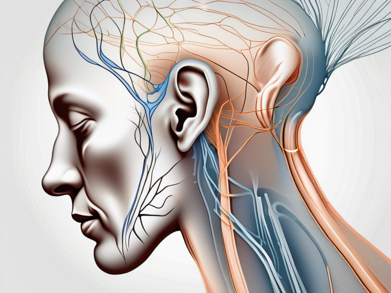
The cochlear nerve, also known as the auditory nerve, holds a critical role in the human auditory system. It is one component of the eighth cranial nerve, alongside the vestibular nerve. The cochlear nerve transmits auditory information from the cochlea, a spiral-shaped structure within the inner ear, to the brain for interpretation and comprehension.
Anatomy of the Cochlear Nerve
The cochlear nerve comprises millions of nerve fibers that originate from the spiral ganglion cells in the cochlea. These nerve fibers bundle together to form the cochlear nerve, which then joins the vestibular nerve to create the eighth cranial nerve.
Within the cochlea, the nerve fibers of the cochlear nerve are organized tonotopically. This means that different frequencies of sound are processed by different regions of the cochlea. The nerve fibers that respond to high-frequency sounds are located near the base of the cochlea, while those that respond to low-frequency sounds are located near the apex.
As the nerve fibers travel from the cochlea towards the brain, they pass through the internal auditory canal, a bony canal within the skull. This canal provides protection to the delicate nerve fibers, ensuring their safe passage towards the brainstem.
The Function of the Cochlear Nerve
The primary function of the cochlear nerve is to transmit auditory signals to the brain. Sound waves are captured by the delicate hair cells lining the cochlea. These hair cells convert the vibrations of sound into electrical signals, which are then relayed through the cochlear nerve to the brainstem and auditory cortex for processing.
Once the electrical signals reach the brainstem, they undergo further processing to extract important information such as sound intensity, pitch, and location. The brainstem acts as a relay station, directing the auditory signals to the appropriate areas of the brain for interpretation.
From the brainstem, the auditory signals are sent to the auditory cortex, which is located in the temporal lobe of the brain. Here, the signals are analyzed and decoded, allowing us to perceive and understand the sounds around us.
Without the functioning of the cochlear nerve, the perception of sound would be severely compromised. Conditions that affect the cochlear nerve, such as acoustic neuroma or damage to the nerve fibers, can result in hearing loss or other auditory impairments.
Research into the cochlear nerve and its role in auditory processing is ongoing. Scientists are continually studying the intricate mechanisms of this nerve to gain a deeper understanding of how we perceive and interpret sound. This knowledge can help in the development of new treatments and interventions for individuals with hearing disorders.
Exploring the Vestibular Nerve
While the cochlear nerve is responsible for auditory perception, the vestibular nerve is integral to maintaining balance and spatial orientation. Working in tandem with the cochlear nerve, the vestibular nerve completes the eighth cranial nerve and contributes to our overall perception of the external world.
Anatomy of the Vestibular Nerve
The vestibular nerve is formed by the nerve fibers originating from the vestibular ganglion, which is located near the inner ear. These fibers extend from the vestibular organs, including the semicircular canals and otolith organs, and merge with the cochlear nerve to create the eighth cranial nerve.
The vestibular ganglion, also known as Scarpa’s ganglion, is a cluster of sensory neuron cell bodies that are responsible for transmitting information from the vestibular organs to the brain. It is situated within the temporal bone, close to the inner ear structures. The ganglion receives signals from the hair cells in the semicircular canals and otolith organs, which detect changes in head position and movement.
The semicircular canals are three fluid-filled structures that are oriented in different planes. They are responsible for detecting rotational movements of the head. Each canal is filled with a gel-like substance called endolymph, and when the head moves, the movement of the endolymph stimulates the hair cells, which then send signals to the vestibular ganglion.
The otolith organs, consisting of the utricle and saccule, are responsible for detecting linear accelerations and changes in head position relative to gravity. These organs contain tiny calcium carbonate crystals called otoliths, which are embedded in a gelatinous substance. When the head moves, the otoliths shift, bending the hair cells and generating electrical signals that are transmitted to the vestibular ganglion.
The Function of the Vestibular Nerve
The primary role of the vestibular nerve is to transmit information regarding head position, movement, and spatial orientation to the brain. This sensory input allows for the regulation of balance, coordination, and posture. By relaying information from the vestibular organs to the brainstem and cerebellum, the vestibular nerve enables us to navigate and interact with the world around us.
When the head moves, the hair cells in the vestibular organs detect the changes in fluid movement and generate electrical signals. These signals are then transmitted through the vestibular nerve to the brainstem, where they are processed and integrated with visual and proprioceptive information. The brainstem, along with the cerebellum, plays a crucial role in coordinating motor responses to maintain balance and posture.
In addition to balance and spatial orientation, the vestibular nerve also contributes to other important functions. It is involved in the vestibulo-ocular reflex, which allows for the coordination of eye movements with head movements. This reflex ensures that the eyes remain focused on a target while the head is in motion, preventing blurring of vision.
The vestibular nerve is also essential for our sense of motion and motion sickness. When there is a mismatch between the signals received from the vestibular system and other sensory systems, such as the visual system, it can result in motion sickness. This occurs when the brain receives conflicting information about motion, leading to symptoms like nausea, dizziness, and vomiting.
Overall, the vestibular nerve plays a vital role in our ability to maintain balance, perceive spatial orientation, and coordinate movements. Without this intricate system, our interactions with the world would be significantly impaired, making the exploration of the vestibular nerve a fascinating and essential area of study.
The Formation of the Eighth Cranial Nerve
We now come to the point where the cochlear nerve and the vestibular nerve unite and form the eighth cranial nerve, also known as the vestibulocochlear nerve. This nerve is responsible for both auditory perception and the maintenance of balance, making it an essential part of the human sensory system.
How the Cochlear and Vestibular Nerves Combine
The merger between the cochlear nerve and vestibular nerve occurs at the internal auditory meatus, a bony canal within the skull. This meatus serves as a passageway for these two nerves to come together and create the eighth cranial nerve. It is fascinating to think about how these distinct nerves, one responsible for hearing and the other for balance, join forces to form a single nerve that plays such a crucial role in our sensory experience.
Once united, these two nerves embark on a remarkable journey within the temporal bone. They traverse through intricate structures, navigating their way towards the brainstem. This journey is not without its challenges, as the nerves must navigate through narrow passages and avoid potential obstructions along the way. It is a testament to the complexity and precision of our anatomy.
The Role of the Eighth Cranial Nerve in Hearing and Balance
As the vestibulocochlear nerve, the eighth cranial nerve assumes the responsibility of transmitting both auditory and vestibular information to the brain. This dual function is truly remarkable, as it allows us to not only hear sounds but also maintain proper balance and orientation in relation to gravity.
When it comes to auditory perception, the vestibulocochlear nerve carries electrical signals from the cochlea, a spiral-shaped structure in the inner ear, to the brain. These signals are then processed and interpreted, allowing us to perceive and understand the rich tapestry of sounds that surround us. From the gentle rustling of leaves to the melodious notes of a symphony, the eighth cranial nerve plays a vital role in our ability to appreciate and enjoy the auditory world.
On the other hand, the vestibular aspect of the eighth cranial nerve is responsible for maintaining our balance and spatial orientation. It receives information from the vestibular organs, which are located within the inner ear and detect changes in head position and movement. This information is then transmitted to the brain, enabling us to navigate our surroundings with ease and grace.
It is important to recognize the significance of the eighth cranial nerve in our daily lives. Dysfunction or damage to this nerve can result in various hearing and balance disorders, such as sensorineural hearing loss or vertigo. These conditions can have a profound impact on an individual’s quality of life, affecting their ability to communicate, enjoy music, or even perform simple tasks that require balance. Understanding the intricate workings of the vestibulocochlear nerve allows us to appreciate the complexity of our sensory system and the importance of its proper functioning.
Disorders Related to the Eighth Cranial Nerve
Unfortunately, the vestibulocochlear nerve is susceptible to a range of disorders. These disorders can have a significant impact on an individual’s auditory perception, balance, and overall well-being. It is crucial to recognize the symptoms of these conditions and seek appropriate medical attention when necessary.
The vestibulocochlear nerve, also known as the eighth cranial nerve, plays a vital role in our ability to hear and maintain balance. This nerve consists of two main components: the cochlear nerve, responsible for transmitting auditory information from the inner ear to the brain, and the vestibular nerve, responsible for transmitting information about balance and spatial orientation.
Disorders related to the eighth cranial nerve can manifest in various ways, affecting different aspects of an individual’s sensory experience. One common symptom is hearing loss, which can range from mild to severe and can have a profound impact on a person’s ability to communicate and engage with their surroundings. Tinnitus, another common symptom, is characterized by a persistent ringing or buzzing sound in the ears. This phantom noise can be incredibly distressing and can significantly impact a person’s quality of life.
Vertigo, a sensation of spinning or dizziness, is also frequently associated with disorders of the vestibulocochlear nerve. This symptom can be debilitating, causing a person to feel off-balance and disoriented. Imbalance, another manifestation of these disorders, can make simple tasks such as walking or standing difficult and increase the risk of falls and injuries.
There are several potential causes of disorders related to the eighth cranial nerve. Trauma, such as a head injury or skull fracture, can damage the nerve and disrupt its normal functioning. Infections, such as meningitis or labyrinthitis, can also affect the nerve and lead to symptoms. Genetic factors can play a role in the development of these disorders, with certain inherited conditions predisposing individuals to auditory and balance problems. Tumors, both benign and malignant, can put pressure on the nerve and interfere with its ability to transmit signals. Additionally, age-related degeneration, known as presbycusis, can contribute to hearing loss and balance issues in older adults.
Diagnosis and Treatment Options
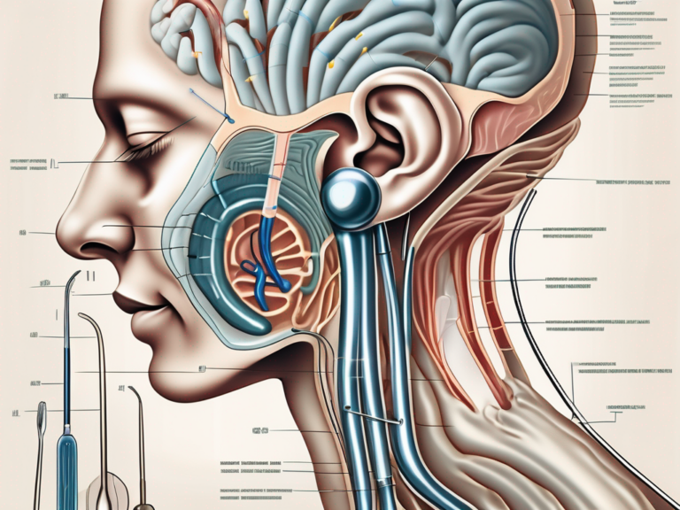
Diagnosing disorders related to the eighth cranial nerve typically involves a combination of medical history evaluation, physical examination, and specialized tests. Audiometry, a hearing test, can assess the extent and nature of hearing loss. Vestibular testing, which includes various assessments of balance and eye movements, can help determine the underlying cause of vertigo and imbalance.
Treatment options for disorders of the eighth cranial nerve depend on the specific condition and its severity. In some cases, medical intervention, such as medications or hearing aids, can help manage symptoms and improve quality of life. For more severe cases or when conservative measures are ineffective, surgical procedures may be necessary. These can range from removing tumors or repairing damaged structures to implanting cochlear or vestibular implants to restore hearing or balance function.
It is crucial for individuals experiencing symptoms related to the vestibulocochlear nerve to consult with a healthcare professional for an accurate diagnosis and appropriate treatment plan. Early intervention can help prevent further deterioration of hearing and balance and improve outcomes.
As we conclude our exploration of the cochlear nerve, the vestibular nerve, and the cranial nerve they form together, it becomes evident how integral these structures are to our hearing, balance, and overall well-being. The intricate connections between the cranial nerves enable us to perceive the world around us, respond to stimuli, and maintain a sense of equilibrium. Through further research, technological advancements, and medical expertise, we continue to improve our understanding of these remarkable neuroanatomical wonders, ultimately enhancing the lives of those affected by disorders related to the eighth cranial nerve.



+ There are no comments
Add yours