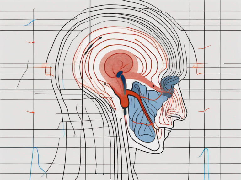

vestibular nerve detects what motion
Uncover the fascinating role of the vestibular nerve in detecting motion and maintaining balance.
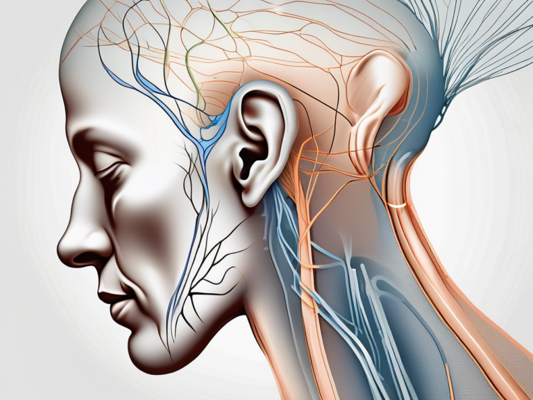
what does the vestibular nerve do in the ear

Discover the fascinating role of the vestibular nerve in the ear and its crucial function in maintaining balance and spatial orientation.
how can you damage vestibular nerve
Learn about the potential causes and consequences of damaging the vestibular nerve in this comprehensive article.
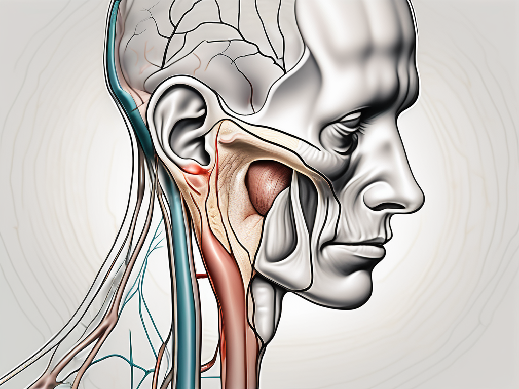
what is labyrinthectomy or vestibular nerve section
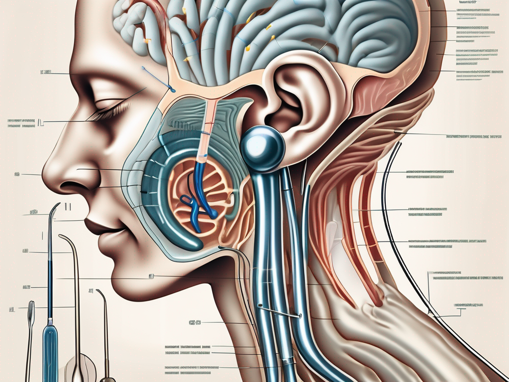
Discover the ins and outs of labyrinthectomy and vestibular nerve section in this comprehensive article.
what is the resting firing potential of the vestibular nerve

Discover the fascinating world of the vestibular nerve’s resting firing potential in this comprehensive article.
how would someone get vestibular nerve damage

Curious about vestibular nerve damage? Discover the causes, symptoms, and potential treatment options in this comprehensive article.
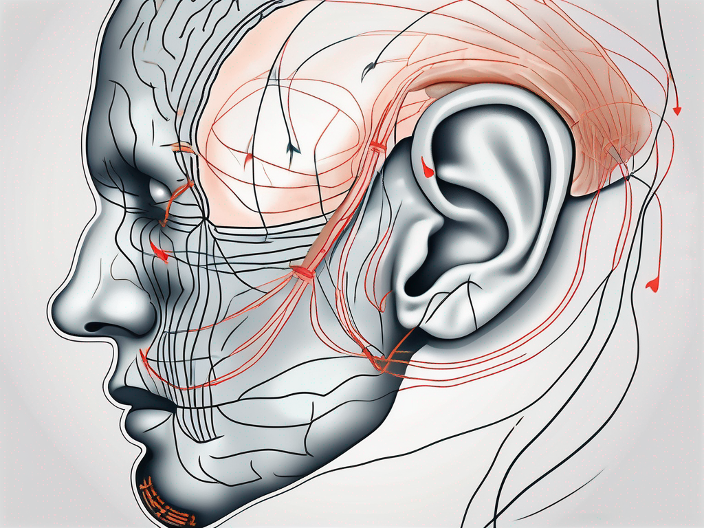
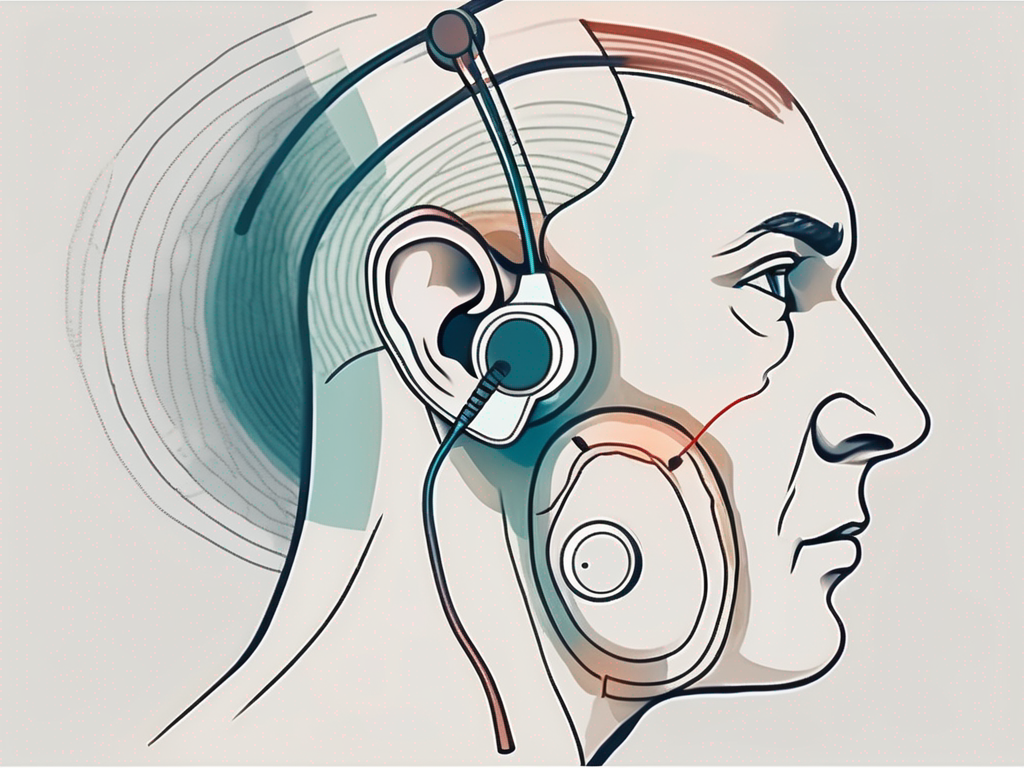
how to stimulate vestibular nerve
Discover effective techniques for stimulating the vestibular nerve and improving your balance and spatial orientation.
what happens when the vestibular nerve is damaged
Discover the effects of a damaged vestibular nerve and how it impacts balance, spatial orientation, and overall quality of life.
why test hearing when vestibular nerve damage is suspected
Discover the importance of testing hearing when suspecting vestibular nerve damage in this insightful article.
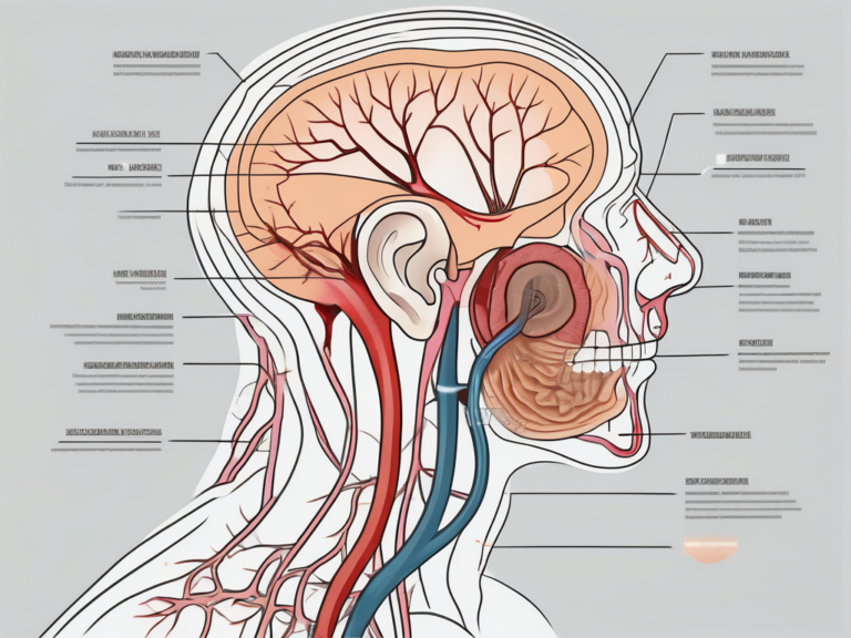
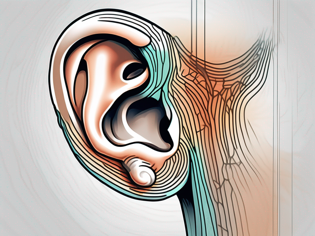
what is the vestibular nerve in the ear
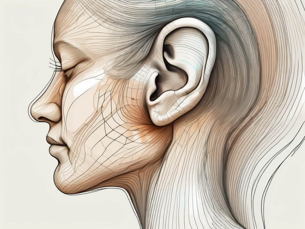
Discover the fascinating role of the vestibular nerve in the ear and its crucial function in maintaining balance and spatial orientation.
You May Also Like:

vestibular nerve detects what motion