The human auditory system is a fascinating and complex system that allows us to perceive and interpret sounds in our environment. Central to this system are two important structures: the cochlea and the vestibular nerve. While they are both integral to our ability to hear and maintain balance, they possess distinct anatomical and functional characteristics that set them apart. In this article, we will explore the anatomy and function of the cochlea and the vestibular nerve, examine their key differences, discuss common disorders that affect them, and delve into the various treatment and management options available.
Understanding the Human Auditory System
Anatomy of the Ear
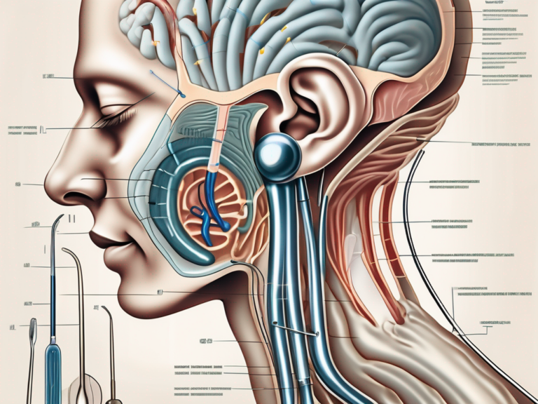
Before diving into the specifics of the cochlea and the vestibular nerve, it is crucial to understand the anatomy of the ear as a whole. The human ear can be divided into three main parts: the outer ear, the middle ear, and the inner ear.
The outer ear consists of the pinna and the ear canal, which work together to collect and funnel sound waves towards the middle ear. The pinna, also known as the auricle, is the visible part of the ear that helps in localizing sound sources. Its unique shape and structure aid in capturing sound waves and directing them into the ear canal.
The ear canal, a narrow tube-like structure, is lined with tiny hairs and glands that produce earwax. These hairs and earwax help to protect the delicate structures of the middle and inner ear from dust, debris, and harmful microorganisms.
Within the middle ear, the sound waves are transformed into mechanical vibrations by the tympanic membrane and three tiny bones known as the ossicles. The tympanic membrane, commonly known as the eardrum, is a thin, cone-shaped membrane that separates the outer ear from the middle ear. When sound waves strike the eardrum, it vibrates, transmitting these vibrations to the ossicles.
The ossicles, consisting of the malleus, incus, and stapes, are the smallest bones in the human body. They form a chain-like structure that amplifies and transmits the vibrations from the eardrum to the inner ear. This mechanical amplification is necessary because the sound waves lose some of their energy when transitioning from air to the fluid-filled inner ear.
These vibrations are then transmitted to the inner ear through the oval window, a membrane-covered opening that connects the middle ear to the cochlea.
The inner ear, nestled deep within the temporal bone, is where the magic of hearing takes place. It is here that the cochlea and the vestibular nerve reside, each playing a vital role in our auditory system.
Function of the Auditory System
The primary function of the auditory system is, of course, hearing. When sound waves reach the cochlea, they are converted into electrical signals that can be interpreted by the brain. This process, known as auditory transduction, is essential for our ability to perceive and comprehend the sounds around us.
The cochlea, a spiral-shaped structure resembling a snail shell, is responsible for converting sound vibrations into electrical signals. It contains thousands of tiny hair cells that are sensitive to different frequencies of sound. When the mechanical vibrations from the ossicles reach the cochlea, they cause the hair cells to bend, triggering the release of chemical signals that generate electrical impulses.
These electrical impulses are then transmitted to the brain via the auditory nerve, where they are processed and interpreted as sound. The brain can distinguish between different frequencies, intensities, and timbres of sound, allowing us to recognize speech, music, and various environmental sounds.
Meanwhile, the vestibular system, which includes the vestibular nerve, is responsible for maintaining our balance and spatial orientation. It detects motion, head position, and gravitational forces, allowing us to adapt to changes in our environment and navigate the world with stability and precision.
The vestibular nerve carries information from the vestibular system to the brain, specifically to the brainstem and cerebellum. This information is crucial for coordinating our movements, maintaining posture, and stabilizing our gaze. It helps us maintain our balance while walking, running, or even standing still, and enables us to make precise adjustments to our body position in response to external stimuli.
In addition to its role in balance, the vestibular system also contributes to our sense of spatial orientation. It allows us to perceive our position in relation to gravity and helps us navigate through space. This is particularly important in situations where visual cues may be limited, such as in the dark or in unfamiliar environments.
In conclusion, the human auditory system is a complex and remarkable system that enables us to perceive and interpret the sounds around us. From the outer ear’s role in collecting sound waves to the inner ear’s conversion of vibrations into electrical signals, every component plays a crucial part in our ability to hear and maintain balance. Understanding the intricate anatomy and function of the auditory system deepens our appreciation for the wonders of human perception.
Deep Dive into the Cochlea
The cochlea, often referred to as the “snail shell,” is a complex spiral-shaped structure that is approximately 3.5 centimeters long. It is a remarkable organ responsible for our sense of hearing, and its intricate design allows us to perceive the world of sound.
Structure of the Cochlea
Let’s take a closer look at the structure of the cochlea. It consists of three fluid-filled compartments, known as scalae, that wind around a central core called the modiolus. These scalae, named the scala vestibuli, scala media, and scala tympani, are interconnected and play a crucial role in the transmission of sound vibrations.
Separating the scalae are two delicate membranes, the Reissner’s membrane and the basilar membrane. The Reissner’s membrane acts as a barrier between the scala vestibuli and the scala media, while the basilar membrane separates the scala media from the scala tympani. These membranes are essential for maintaining the integrity of the cochlear compartments and ensuring the proper transmission of sound.
Lining the basilar membrane are specialized sensory cells called hair cells. These remarkable cells contain tiny hair-like structures called stereocilia. The hair cells are arranged in rows along the length of the cochlea, with different regions responding to different frequencies of sound. The stereocilia of the hair cells are bathed in a fluid called endolymph, which plays a crucial role in the transduction of sound vibrations into electrical signals.
Role of the Cochlea in Hearing
The cochlea’s primary function is to transform sound vibrations into electrical signals that can be sent to the brain. This intricate process begins when sound waves enter the cochlea through the oval window, a membrane-covered opening in the bony labyrinth of the inner ear.
As the sound vibrations travel through the fluid-filled cochlear compartments, they cause the basilar membrane to vibrate. The basilar membrane is not uniform in its structure; it varies in width and stiffness along its length. This variation allows different regions of the basilar membrane to respond to different frequencies of sound. When a specific frequency of sound reaches the cochlea, it causes the corresponding region of the basilar membrane to vibrate most vigorously.
These vibrations stimulate the hair cells that are located above the specific region of the basilar membrane responding to the frequency of the sound. The stereocilia on the hair cells bend in response to the movement of the basilar membrane, initiating a cascade of biochemical events within the hair cells.
As the stereocilia bend, they open ion channels, allowing ions to flow into the hair cells. This influx of ions generates electrical signals, which are then transmitted to the brain through the auditory nerve fibers. The auditory nerve carries these electrical signals to the auditory cortex in the brain, where they are processed and interpreted as sounds.
The cochlea’s ability to discriminate between different frequencies of sound is a remarkable feat. It is thanks to the precise arrangement of the hair cells along the basilar membrane and the specific regions of the cochlea that respond to different frequencies. This tonotopic organization allows us to perceive a wide range of sounds, from the low rumble of thunder to the high-pitched notes of a songbird.
Understanding the structure and function of the cochlea provides us with insights into the incredible complexity of our auditory system. The cochlea’s intricate design and its role in transforming sound vibrations into electrical signals highlight the remarkable capabilities of the human ear.
Exploring the Vestibular Nerve
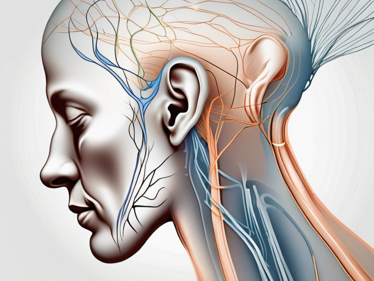
The vestibular nerve, also known as the vestibulocochlear nerve or the eighth cranial nerve, is a paired nerve that consists of two branches: the cochlear branch and the vestibular branch.
The cochlear branch is responsible for transmitting auditory information from the cochlea to the brain. It allows us to perceive and interpret sounds, enabling us to enjoy music, communicate with others, and be aware of our environment.
On the other hand, the vestibular branch carries signals related to balance and spatial orientation. It plays a crucial role in maintaining our equilibrium and coordinating our movements.
Anatomy of the Vestibular Nerve
The vestibular nerve originates from the vestibular ganglion, located within the inner ear. This ganglion is a cluster of nerve cell bodies that serve as the starting point for the vestibular branch of the nerve.
From the vestibular ganglion, the vestibular nerve travels through the internal auditory canal, which is a bony canal within the temporal bone of the skull. This canal provides a protected pathway for the nerve fibers as they make their way towards the brainstem.
Once inside the brainstem, the vestibular nerve connects to specific regions that are responsible for processing and integrating the signals it carries. These regions include the vestibular nuclei, which are located deep within the brainstem and play a vital role in maintaining balance and coordinating eye movements.
Function of the Vestibular Nerve in Balance
The vestibular nerve plays a crucial role in maintaining balance and spatial awareness. It detects the position and movement of the head, allowing us to adjust our posture and coordinate our movements accordingly.
When we move our head, the fluid-filled canals of the inner ear, known as the semicircular canals, are set in motion. This movement stimulates specialized sensory cells within the canals, which then send signals to the vestibular nerve.
The vestibular nerve carries these signals to the brain, specifically to the vestibular nuclei. The brain processes this information and generates appropriate responses to maintain balance and stability.
By sending signals to the brain about changes in head position or motion, the vestibular nerve helps ensure that we remain stable and oriented in relation to gravity and our surroundings. It allows us to walk, run, jump, and perform various activities without losing our balance.
In addition to maintaining balance, the vestibular nerve also contributes to other important functions. It helps us maintain a sense of spatial orientation, allowing us to navigate our environment effectively. It also plays a role in coordinating eye movements, ensuring that our gaze remains stable even when our head is in motion.
In summary, the vestibular nerve is a remarkable component of our nervous system. It enables us to perceive sound, maintain balance, and navigate our surroundings. Without this intricate network of nerve fibers, our ability to interact with the world around us would be greatly impaired.
Key Differences between the Cochlea and Vestibular Nerve
Structural Differences
One of the main differences between the cochlea and the vestibular nerve lies in their structure. The cochlea, as previously mentioned, is a spiral-shaped structure that is dedicated to the processing of sound vibrations. It is a remarkable organ that resembles a snail shell and is located in the inner ear. The cochlea is composed of three fluid-filled chambers, known as the scala vestibuli, scala media, and scala tympani. These chambers are separated by delicate membranes and contain specialized sensory cells called hair cells. These hair cells are responsible for converting mechanical vibrations into electrical signals that can be transmitted to the brain for interpretation.
In contrast, the vestibular nerve is a branch of the eighth cranial nerve that is responsible for transmitting information related to balance and spatial orientation. It is a complex network of nerve fibers that connects the inner ear to the brain. The vestibular nerve consists of two main components: the superior vestibular nerve, which carries signals related to head movement and rotation, and the inferior vestibular nerve, which transmits signals related to linear acceleration and gravity. These two components work together to provide the brain with essential information about the body’s position and movement in space.
Functional Differences
Another significant difference between the cochlea and the vestibular nerve lies in their respective functions. The cochlea’s primary role is to convert sound vibrations into electrical signals that can be interpreted by the brain, thus enabling us to hear. When sound waves enter the ear, they cause the eardrum to vibrate. These vibrations are then transmitted through the middle ear to the cochlea, where they stimulate the hair cells. The hair cells respond to different frequencies of sound and send corresponding electrical signals to the brain via the auditory nerve. The brain then processes these signals, allowing us to perceive and understand sounds.
On the other hand, the vestibular nerve plays a vital role in balance regulation by transmitting signals related to head position, motion, and gravitational forces. It provides the brain with constant updates about the body’s orientation in space, allowing us to maintain our balance and coordinate our movements. When we move our head or change our body position, the vestibular system detects these changes and sends signals to the brain, which then adjusts our posture and activates the appropriate muscles to keep us stable. Without the vestibular nerve, simple tasks like walking, running, or even standing upright would be extremely challenging.
In addition to balance regulation, the vestibular nerve also contributes to other important functions, such as eye movement control and spatial awareness. It works in conjunction with other sensory systems, including vision and proprioception, to provide a comprehensive understanding of our body’s position and movement in relation to the environment.
Common Disorders Affecting the Cochlea and Vestibular Nerve
Cochlear Disorders and Their Impact on Hearing
Disorders affecting the cochlea can have a profound impact on an individual’s ability to hear. Conditions such as sensorineural hearing loss, noise-induced hearing loss, and Meniere’s disease can all affect the functioning of the cochlea and result in varying degrees of hearing impairment. It is essential for individuals experiencing any changes in their hearing to seek medical evaluation and treatment promptly.
Vestibular Nerve Disorders and Their Effect on Balance
Disorders affecting the vestibular nerve can lead to problems with balance and spatial orientation. Conditions such as vestibular neuritis, benign paroxysmal positional vertigo (BPPV), and labyrinthitis can all disrupt the normal functioning of the vestibular nerve and cause dizziness, vertigo, and problems with balance. Individuals experiencing these symptoms should consult with a healthcare professional for proper diagnosis and treatment.
Treatment and Management of Cochlear and Vestibular Nerve Disorders
Medical Interventions
The treatment and management of cochlear and vestibular nerve disorders depend on the specific condition and its underlying cause. Medical interventions may include medications to alleviate symptoms, surgical procedures to restore or improve hearing, or interventions aimed at addressing the root cause of the disorder. It is crucial for individuals with cochlear or vestibular nerve disorders to consult with medical professionals experienced in these areas to determine the most appropriate course of action.
Rehabilitation and Therapy Options
In addition to medical interventions, rehabilitation and therapy options are often recommended to individuals with cochlear and vestibular disorders. These options may include auditory training, balance exercises, strategies for managing symptoms, and counseling to address the emotional and psychological impact of hearing and balance impairments. Comprehensive care that combines medical interventions with rehabilitative support can provide individuals with a holistic approach to managing their conditions and improving their quality of life.
Conclusion
In conclusion, the cochlea and the vestibular nerve are two distinct structures within the human auditory system that serve essential but separate functions. While the cochlea is responsible for processing sound vibrations and enabling us to hear, the vestibular nerve plays a crucial role in regulating balance and spatial orientation. Understanding the differences between these structures is vital in diagnosing and managing disorders that affect hearing and balance. If you are experiencing any symptoms related to your hearing or balance, it is imperative to seek consultation with a healthcare professional who specializes in the auditory system and vestibular function. With proper diagnosis and treatment, individuals can regain or maintain optimal functionality in these crucial aspects of human experience.



+ There are no comments
Add yours